




Key Takeaways
- MIT CSAIL’s xvr pretrains on 2,000+ whole-body scans, then fine-tunes to a new patient in about five minutes with sub-millimeter 2D/3D registration accuracy.
- It synthesizes roughly 1,000 physics-simulated X-ray projections per second from the patient’s own CT or MRI, avoiding generative hallucination of anatomy.
- A generalist challenger, XPos3R, claims sub-4 mm 3D error with no patient-specific tuning, but that preprint result is not yet independently replicated.
Table of Contents
Sub-Millimeter AI Registration Arrives for Surgical X-Rays
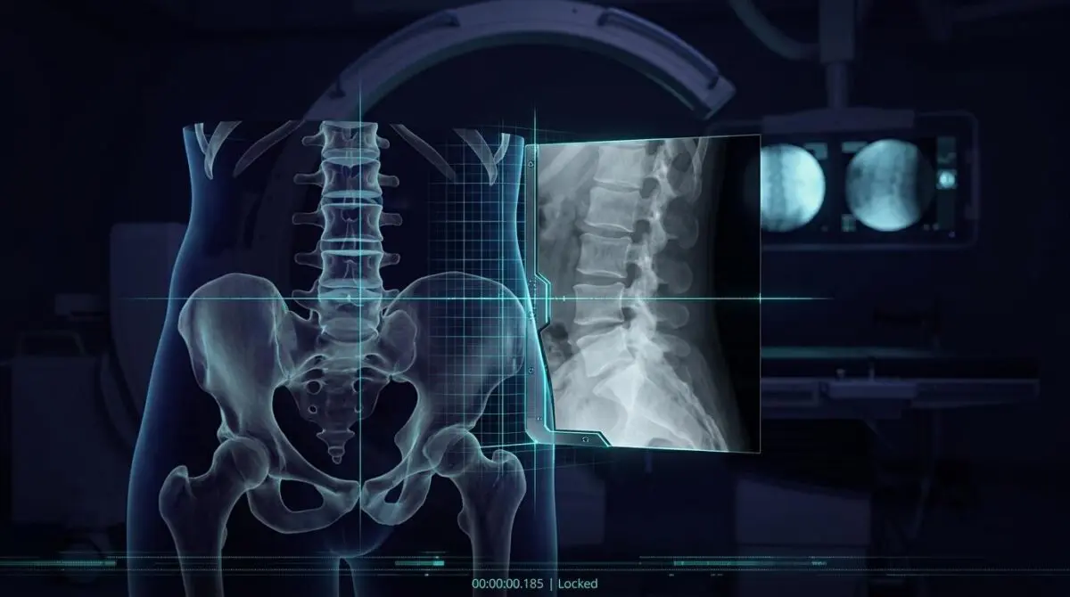
MIT News broke the story today on a patient-specific AI model called xvr that can register intraoperative flat X-ray frames against preoperative 3D CT or MRI volumes within seconds.
The technique, developed at MIT CSAIL with collaborating institutions, achieves sub-millimeter image alignment and delivers an order-of-magnitude improvement over prior AI registration tools.
Published today in Nature, the work targets a bottleneck that makes minimally invasive and emergency surgical navigation slower and less precise.
As MIT News frames it, this is not a narrow academic result.
It changes whether hospitals can safely guide catheters, endoscopes, and robotic instruments through the body using flat real-time X-ray images.
From Physics-Simulated X-Rays to a Five-Minute Patient Model
Clinicians performing minimally invasive procedures often rely on high-speed mobile X-ray scanners to produce flat, real-time images from any angle.
To determine where a tool sits relative to critical anatomy, they manually align those projections with a preoperative CT or MRI scan.
That registration step is slow, subjective, and highly dependent on the clinician’s ability to read grainy 2D images.
It takes decades of training for a clinician to become skilled enough to see grainy, 2D images and understand how everything is oriented. We want to make these 2D X-rays more informative, so it becomes safer and easier to do these life-saving procedures.
Lead author Vivek Gopalakrishnan, a postdoc in MIT CSAIL, frames the problem as one of making flat X-rays carry spatial intelligence.
xvr attacks the issue differently from standard one-size-fits-all AI models.
Instead of training a single model that generalizes across every patient anatomy, the team builds a model tailored specifically to each patient.
The system starts with a patient’s preoperative 3D scan and synthesizes about 1,000 realistic X-ray projections every second across many viewing angles.
It relies on a physics simulation of how X-rays interact with tissue, rather than on generative image hallucination.
Because the synthetic data derive directly from that patient’s own CT or MRI, the model has no room to invent anatomical structure.
Building a registration model from zero for each patient would require roughly 12 hours, making emergency use impossible.
To solve that, the researchers pretrained a foundation model using whole-body scans from more than 2,000 individuals representing different ages, imaging modalities, and anatomical regions.
That pretrained model fine-tunes to a new patient in roughly five minutes and matches the accuracy of a model trained from zero.
- 2,000+ whole-body scans used to pretrain the foundation model.
- 1,000 synthetic X-ray projections per second generated from the patient’s own scan.
- 5 minutes to adapt to a new patient.
- Sub-millimeter accuracy on real 2D/3D registrations from five hospitals.
The evaluation used real 2D-to-3D image pairs gathered from five hospitals, spanning multiple bones and organ systems across both adult and pediatric patients.
The method was more accurate and more robust than other AI-based approaches, and it still ran quickly enough for emergency use.
Clinical Validation and the Patient-Specific vs. Generalist Race
A prospective pilot study published in JoVE offers an early read on AI-augmented navigation in a live operating room.
Researchers enrolled 14 patients over a one-year window ending in December 2024 to evaluate the LungVision system for intraoperative pulmonary nodule localization.
Every target lesion was localized, all resections were complete, and no localization-related complications were recorded.
Mean navigation time was 38.6 minutes, and 71.4 percent of nodules were pathologically malignant.
The study authors caution that larger trials are needed, but the feasibility signal is clear: AI-assisted navigation can fit into existing surgical workflows without major infrastructure changes.
A September 2026 arXiv preprint adds sharper competitive pressure.
The paper, accepted to ECCV 2026, introduces XPos3R, a cross-modal transformer designed to eliminate preoperative preparation entirely.
The authors report that one pretrained XPos3R model beat patient-specific methods on both accuracy and robustness in real-world benchmarks.
With only seconds of test-time optimization, the preprint reports sub-4 mm 3D error and sub-1 mm reprojection error.
That claim remains preprint-level and has not yet been independently replicated at production scale.
But the strategic implication is direct: if a generalizable model can match patient-specific accuracy without the five-minute adaptation window, the deployment calculus for surgical robotics shifts.
A separate ACM Digital Library study extends the same direction in spine surgery, using a coarse-to-fine Shape-aware and Fine-focused Transformer to fuse intraoperative ultrasound with preoperative CT.
That work reports state-of-the-art registration accuracy on public datasets and on an author-built clinical vertebra dataset, while noting that lightweight models and non-rigid registration remain open problems.
The xvr team is already collaborating with surgical robotics companies and clinical groups to move from validation toward deployment.
That timing matters because the field is now splitting into two architectures: patient-specific tuning versus generalized pretraining.
xvr anchors the patient-specific side with peer-reviewed sub-millimeter results and a five-minute adaptation window.
XPos3R represents the generalist challenger, betting that a single model can erase even that short delay.
What neither approach yet has is large-scale prospective surgical outcome data.
LungVision’s feasibility study shows the clinical route is viable, but its 14-patient sample is far from definitive.
When Surgical Navigation Becomes a Software Problem
The near-term consequence is not that AI will replace the surgeon, but that surgical navigation is becoming a software problem with patient-specific accuracy.
For teams building AI validation pipelines that must separate peer-reviewed results from preprint claims, Andres SEO Expert’s programmatic SEO AI automation is built to surface the right technical audience — contact Andres SEO Expert.
Frequently Asked Questions
What is xvr and what does it do?
xvr is a patient-specific AI model developed at MIT CSAIL that registers intraoperative flat 2D X-ray frames against preoperative 3D CT or MRI volumes within seconds. It achieves sub-millimeter image alignment and is designed to make minimally invasive and emergency surgical navigation faster and more precise.
How accurate is AI registration for surgical X-rays?
The xvr method achieves sub-millimeter accuracy on real 2D-to-3D registrations from five hospitals, spanning multiple bones and organ systems in adult and pediatric patients. The researchers report an order-of-magnitude improvement over prior AI registration tools while still running quickly enough for emergency use.
Why is patient-specific AI used instead of a general model?
Patient-specific AI builds a model tailored to each patient from their own preoperative CT or MRI. The system uses a physics simulation of X-ray tissue interaction to synthesize about 1,000 realistic projections per second, so it has no room to invent anatomical structure. A pretrained foundation model then makes adaptation practical.
How long does it take to adapt xvr to a new patient?
After pretraining on whole-body scans from more than 2,000 individuals, xvr fine-tunes to a new patient in roughly five minutes. Building a registration model from zero for each patient would take about 12 hours, which would make emergency use impossible.
Has AI-augmented surgical navigation been tested in real operations?
A JoVE prospective pilot study evaluated the LungVision system in 14 patients over a one-year window ending in December 2024. Every target lesion was localized, all resections were complete, and no localization-related complications were recorded. Mean navigation time was 38.6 minutes, but larger trials are still needed.
What is XPos3R and how does it challenge patient-specific AI?
XPos3R is a cross-modal transformer introduced in a September 2026 arXiv preprint accepted to ECCV 2026. It aims to eliminate preoperative preparation entirely, and the authors report that one pretrained model beat patient-specific methods with seconds of test-time optimization, reaching sub-4 mm 3D error and sub-1 mm reprojection error. Those claims remain preprint-level and have not been independently replicated at production scale.
Is AI replacing surgeons in minimally invasive surgery?
No. The near-term consequence is that surgical navigation is becoming a software problem with patient-specific accuracy. AI can help guide catheters, endoscopes, and robotic instruments through the body using real-time X-ray images, but the surgeon remains central and neither patient-specific nor generalist approaches yet have large-scale prospective surgical outcome data.